Annotated Bibliography
Sequencing ancient calcified dental plaque shows changes in oral microbiota with dietary shifts of the Neolithic and Industrial revolutions
Adler CJ, Dobney K, Weyrich LS, Kaidonis J, Walker AW, et al. 2013. Sequencing ancient calcified dental plaque shows changes in oral microbiota with dietary shifts. Proc Natl Acad Sci USA. 110(15):5647–5652. doi:10.1073/pnas.1305477110. PMCID: PMC3996550.
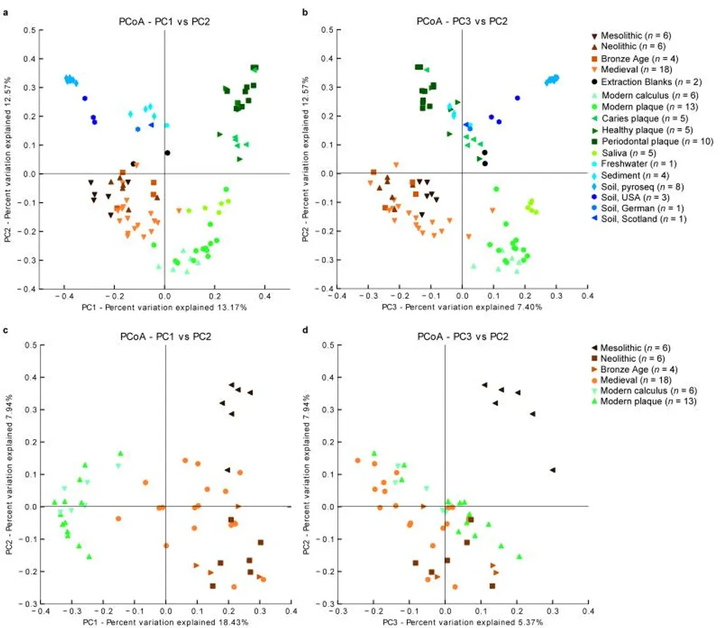
In this paper, researchers study ancient dental calculus to examine how changes in human dietary habits affected the composition of mouth bacteria. They compare hunter-gatherers, early farmers, and industrial people. During the Industrial Revolution, cariogenic bacteria became dominant. Apparently, as the diet contains more starch and sugar, the oral microbiota shifts to a more cariogenic profile.
I use Figure 2 to show how different lifestyles are associated with different mouth bacteria at different times. Ancient calculus from hunter-gatherer and early farming groups is very different compared with modern plaque. When the diet started to added more sugar, people tend to developed more dental problems. That is one reason caries has increased in recent times. I think it's a good idea to use this article as a starting point to discuss a common dental issue: dental plaque. Then I will link it to my main topic (third molars): the same diet shift that changed the biofilm also reduced chewing demand, which is part of why many modern jaws are smaller and leave less space for M3.
A dental revolution: The association between occlusion and chewing behaviour
Silvester CM, Kullmer O, Hillson S. A dental revolution: The association between occlusion and chewing behaviour. PLoS One. 2021 Dec 15;16(12):e0261404. doi: 10.1371/journal.pone.0261404. PMID: 34910787; PMCID: PMC8673603.
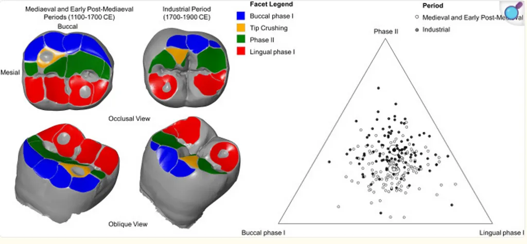
This article explains how the modern diet, rich in soft foods, and certain oral habits, such as chewing food less thoroughly, can affect jaw development and dental occlusion. Chewing less during childhood can lead to smaller dental arches, resulting in less space for the teeth. The post-industrial diet also had negative effects, as foods became increasingly processed and standardized, containing more and more sugar, thus contributing to the emergence of new dental problems seen today. The article supports the idea that changes in food texture (and eating frequency/snacking habits) contribute to the increased prevalence of crowded teeth and problems with wisdom teeth in current patients.
I plan to use this paper as an introduction to the topic I will focus on: how, at the beginning of the industrial era, numerous dental problems began to emerge, from tooth decay to changes in jaw structure due to alterations in food texture and the resulting changes in chewing habits.
Third Molar Agenesis Is Associated with Facial Size
Gkantidis N, Fan J, Xiong H, Halazonetis DJ, Vasilakos G. 2021. Third molar agenesis is associated with facial size. Biology. 10(7):650.
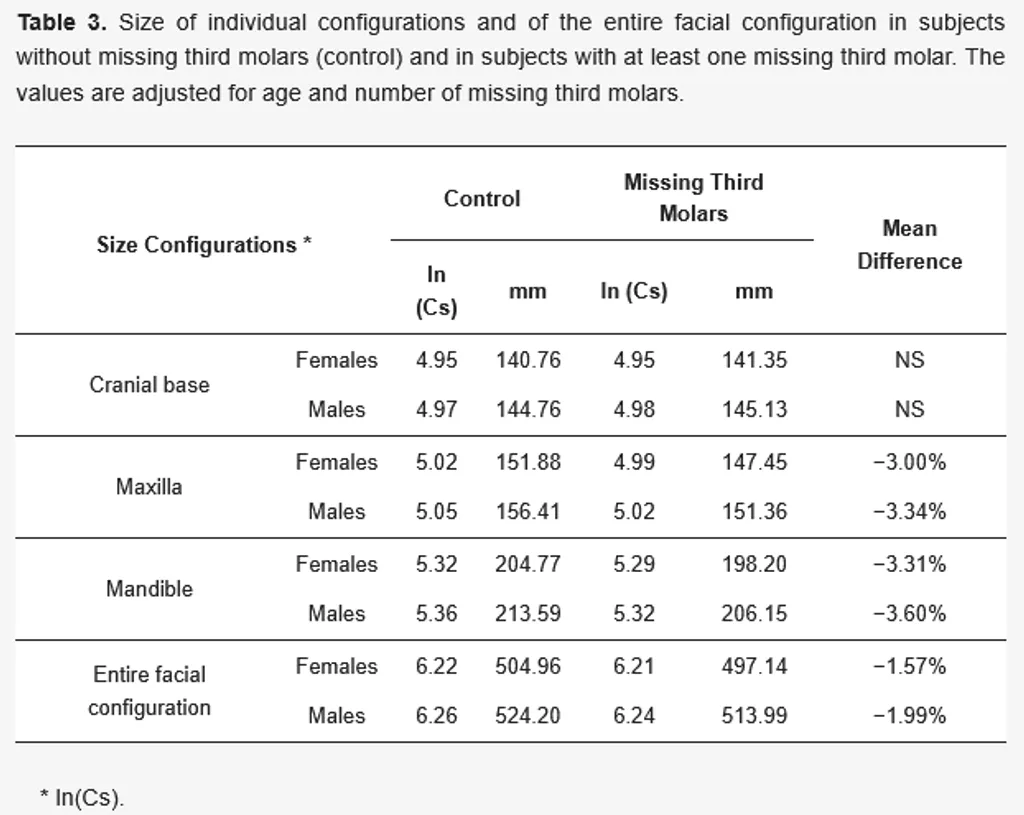
This article analyzes the relationship between the absence of third molars (M3) and smaller jaw and facial dimensions. Using geometric morphometric, the authors compare patients with and without agenesis of the third molars. The results showed that when one or more third molars are missing, the maxilla and mandible are smaller, and the facial structure is also smaller; this effect intensifies as the number of missing third molars increases. The cranial base does not show this effect. Furthermore, this association is observed even when the agenesis affects only one dental arch. Therefore, it is concluded that the agenesis (absence) of third molars is related to smaller jaws and less space.
For my project, I will use Table 3 from the article, which shows the differences in the size of the maxilla, mandible, and face in men and women with and without third molars. This table will be very useful for explaining why many patients today have insufficient posterior space and problems with the eruption of their third molars.
Third molar agenesis in modern humans with and without agenesis of other teeth
Scheiwiller M, Oeschger ES, Gkantidis N. 2020. Third molar agenesis in modern humans with and without agenesis of other teeth. PeerJ 8:e10367.
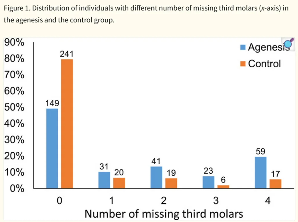
This article discusses the frequency of missing third molars (M3) and its relationship with the absence of other teeth. The authors compare two groups: (1) individuals with agenesis of some teeth (excluding M3s) and (2) individuals without dental agenesis. The results show that M3 agenesis is common and more frequent in the group with other dental anomalies. Differences according to sex and jaw (upper and lower) are also analyzed.
I will use the graph found in the article because it compares two groups: people with agenesis of other teeth and a control group. It shows how many third molars are missing (from 0 to 4) in each person. In the group with agenesis, the absence of all four third molars is more common, while in the control group, the absence of third molars is less frequent.
Impaction of lower third molars and their association with age: radiological perspectives
Ryalat S, Dar-Odeh N, Abul Hasan R, Alrwaily M, Baqain ZH. 2018. Impaction of lower third molars and their association with age. BMC Oral Health. 18:58.
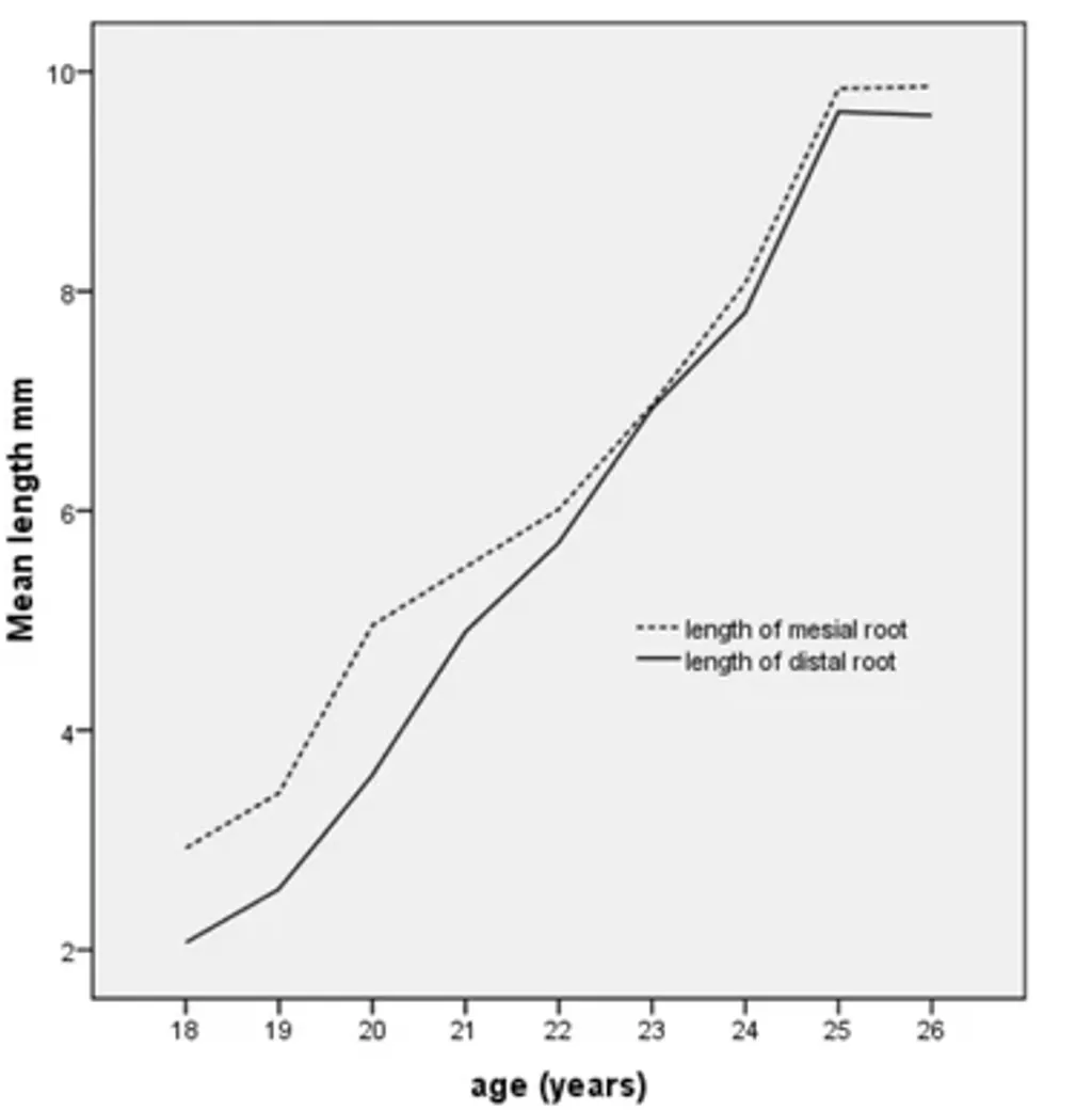
This article analyzes numerous panoramic radiographs of young adults, focusing on impacted lower third molars. It describes the most common angulations (mesioangular impaction is very frequent) and how these characteristics vary with age. As people age, vertical impaction becomes more common, the retromolar space tends to increase, and root formation continues.
I plan to include Figure 3 because it illustrates how the root size of the lower third molar increases with age; after the age of 20, the root continues to grow, which can make extraction more difficult.
Evolution of the oral microbiome and dental caries
Adler, C. J., Browne, G. V., Sukumar, S., & Hughes, T. (2017). Evolution of the oral microbiome and dental caries. Current Oral Health Reports, 4(4), 264–269. https://doi.org/10.1007/s40496-017-0151-1
The article reviews and synthesizes genetic evidence showing how transition to agriculture transformed the oral microbiome from a diverse state to a low-diversity dominated by acid producing pathogens. The article links dietary carbohydrates to the selective pressure of streptococcus mutants. Mainly the modern diets allowed the growth of S. mutants. This was achieved by analyzing fossil tooth evidence.
This paper was utilized to highlight how the change in diet led to the constant evolution of our oral microbiomes. This helps put a good picture in the evolution of tooth. This bring some questions such as: Is a less diverse microbiome good? Usually diversity helps with better evolutionary traits and survival.
Evolutionary and population genomics of the cavity causing bacteria Streptococcus mutans
Cornejo, O. E., Lefébure, T., Bitar, P. D. P., Lang, P., Richards, V. P., Eilertson, K., Do, T., Beighton, D., Zeng, L., Ahn, S.-J., Burne, R. A., Siepel, A., Bustamante, C. D., & Stanhope, M. J. (2013). Evolutionary and population genomics of the cavity causing bacteria Streptococcus mutans. Molecular Biology and Evolution, 30(4), 881–893. https://doi.org/10.1093/molbev/mss278
This article uses genome sequencing of S. mutants strains to reconstruct its evolutionary history. The main findings reveal that exponential growth began around 10 thousand years ago, coinciding with the beginning of agriculture. Other findings revealed that genes under strong pressure including acid tolerance, enabled S. mutants to exploit carbohydrate rich diets.
This paper was useful as a support for the Adler et al. paper. Mainly because this paper looks at the other side of the coin. The exponential growth of S. mutants led to the human body to adapt. Additionally, I will use figure 1 of this paper because it visualizes the evolution and growth of S. mutant
Exploiting the oral microbiome to prevent tooth decay: Has evolution already provided the best tools?
Baker, J. L., & Edlund, A. (2019). Exploiting the oral microbiome to prevent tooth decay: Has evolution already provided the best tools? Frontiers in Microbiology, 9, Article 3323. https://doi.org/10.3389/fmicb.2018.03323
This paper explores how evolution shaped a colonization-resistant oral microbiome that naturally suppresses cariogenic pathogens such as the one explored in this page the S. mutants. The author details dental plaque showing the development of different colonizers when they arrived to the teeth. For instance, the early colonizers such as S. sanguinis fought with S. mutants but in the end S. mutants dominated via acid production. The focus is mainly to argue against normal interventions (e.g., fluoride) to something more ecologybased such as arginine prebiotics.
This paper is relevant in this research as the final puzzle of the story. While the other two papers about this topic talk about microbes that fight colonizers (such as S. mutant). This paper focuses mostly on the restoration process of the teeth. This means that evolution has in a sense weakened our teeth and is calling for a path that can allow us to reverse the diet-induced dental decay (S. mutant dominance). This comes back to the question of: Is a less diverse microbiome in the teeth good? This paper answers the question with a no and is calling for a way to reverse this part in evolution.
Copper and zinc isotope ratios in human bone and enamel. American Journal of Physical Anthropology
Jaouen, K., Herrscher, E., & Balter, V. (2017). Copper and zinc isotope ratios in human bone and enamel. American Journal of Physical Anthropology, 164(3), 407–421. https://doi.org/10.1002/ajpa.23132
This paper examines how zinc and copper isotope ratios in human enamel and bone can reveal long-term changes in diet across different historical periods. The researchers analyzed samples from three populations that lived in the 17th–18th century, the 19th century, and modern times. By comparing the isotopic signatures in enamel, the scientists could determine how dietary habits shifted over time. Results show that humas have lower Zn compared to earlier populations whose diets contained less meat.
Figure 5 in this paper compares Zn values in historic and modern humans, showing clear differences between earlier populations (St-Laurent and CCEC) and modern individuals. This graph is helpful because it visually demonstrates that enamel retains a dietary fingerprint unique to each period. Modern samples cluster at lower isotope values, while earlier populations show higher values, emphasizing that shifts in diet created these differences.
Magnesium isotopic composition of modern human teeth enamel and its implications for dietary reconstructions
Gao, F., Zhang, P., Liu, K., Ling, X., & Huang, K.-J. (2023). Magnesium isotopic composition of modern human teeth enamel and its implications for dietary reconstructions. Frontiers in Earth Science.
This paper investigates how magnesium isotopes in human enamel reflect different cerealbased diets in modern Chinese populations. The researchers sampled enamel from individuals living in four regions of China, each with distinct staple foods such as rice in the south and wheat in the north. Using magnesium isotope analysis, they examined how much Mg was incorporated into enamel and whether isotope patterns corresponded to local diets. Their results show that people from rice-eating regions had higher Mg concentrations and heavier Mg values in their enamel, while people from wheat-eating northern regions had lighter values. The study ruled out environmental and physiological explanations and concluded that these enamel differences are caused by diet alone.
Figure 2 in the paper shows a strong linear relationship between the magnesium provided by local crops and the magnesium found in human enamel. This figure is important because it clearly demonstrates that staple foods, not environmental background levels, control enamel Mg content. As the proportion of rice increases, enamel Mg becomes heavier, while higher wheat intake produces lighter values